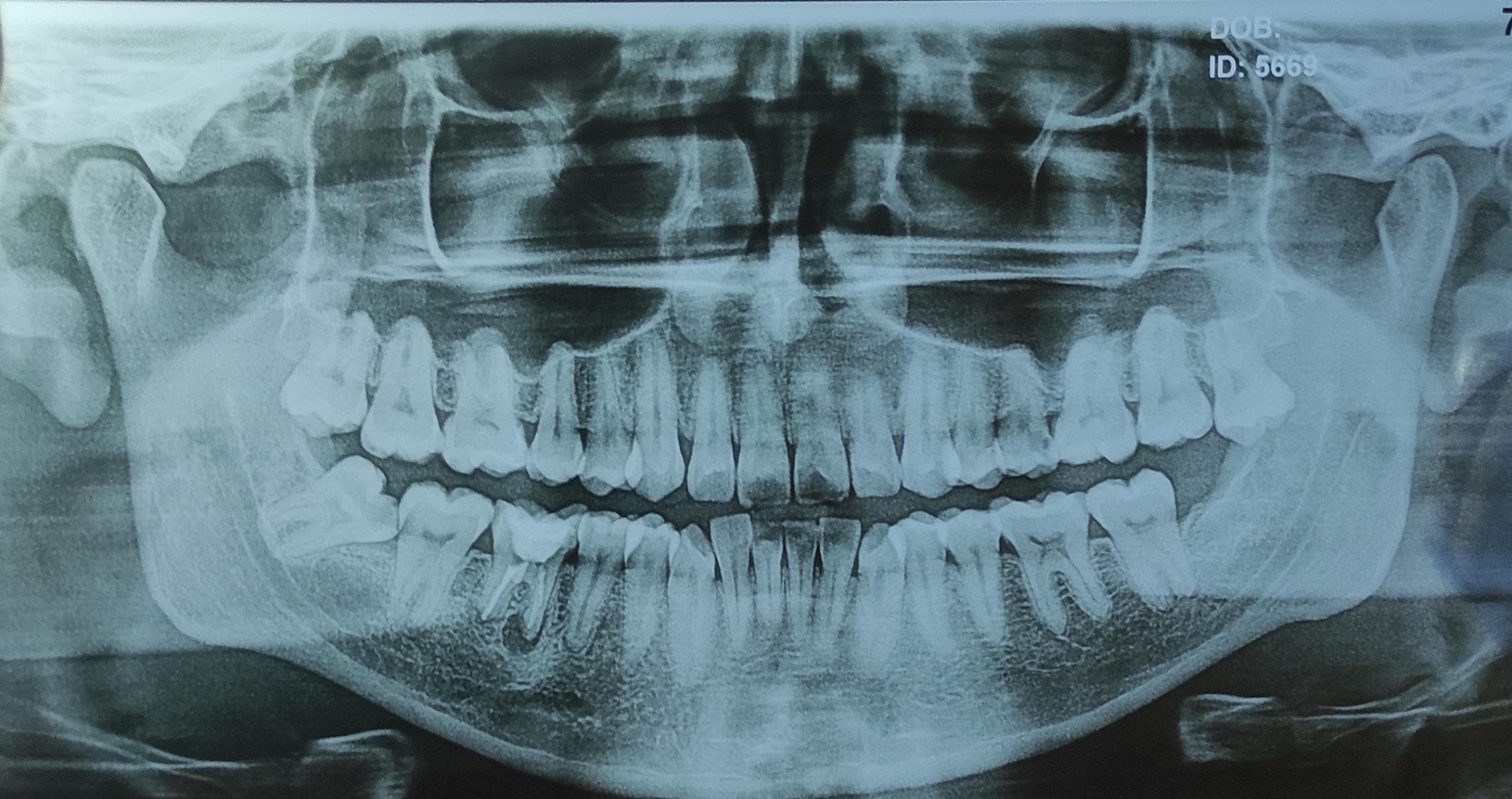
If you’ve been told you need wisdom teeth surgery, your impacted wisdom tooth x-ray (often an OPG scan) is the roadmap your oral surgeon uses to avoid nerves, plan extractions, and prevent surprises. In this guide, you’ll see real annotated x-rays, learn how to spot risks, and understand why 92% of surgeons insist on OPG scans
Why X-Rays Are Non-Negotiable for Impacted Wisdom Teeth
- Depth of impaction (fully buried vs. partially erupted)
- Root shape & nerve proximity (avoiding permanent numbness)
- Hidden damage to neighboring teeth
- Cysts/tumors (3-5% of cases)
Types of Impacted Wisdom Teeth on X-Ray (With Visuals)
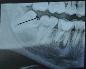
🔍 1. Mesial Impaction (Most Common)
- X-ray sign: Tooth angled forward toward 2nd molar
- Risks: Cavities in adjacent tooth, crowding
- Surgery difficulty: Moderate
🔍 2. Vertical Impaction
- X-ray sign: Tooth upright but trapped under gums
- Risks: Pericoronitis (infection), gum disease
- Surgery difficulty: Low
🔍 3. Horizontal Impaction (Most Complex)
- X-ray sign: Tooth sideways, 90° to other teeth
- Risks: Root damage to 2nd molar, jaw fractures
- Surgery difficulty: High
🚨 Nerve Proximity on X-Ray: Avoiding Permanent Numbness
Your OPG scan shows the inferior alveolar nerve (IAN) – damage causes lip/chin numbness. Red flags:
- Dark band touching root on x-ray
- Root curvature hooking around nerve
- No gap between tooth and nerve canal
Also Read: NHS Wisdom Tooth Removal Risks
How Surgeons Plan Extraction Using Your X-Ray
Step 1: CBCT Scan for High-Risk Cases
If nerve contact is suspected, a 3D CBCT scan provides millimeter-precision:
- Reduces nerve injury risk by 83% (IJOMS 2025)
- Cost: Typically $150-$350
Step 2: Surgical Approach Mapping
- Incision points avoiding nerves
- Bone removal zones (yellow on your x-ray)
- Tooth sectioning plan (cutting tooth to remove safely)
5 Questions to Ask After Seeing Your Impacted Wisdom Tooth X-Ray
- “Is the nerve canal touching my tooth root?”
- “Will you need to remove bone?”
- “Could my second molar be damaged?”
- “Is there a cyst visible?”
- “Do I need a 3D scan for safety?”
Impacted Wisdom Teeth Complications Seen on X-Ray
| Complication | X-Ray Sign | Treatment Urgency |
|---|---|---|
| Dentigerous Cyst | Dark bubble around crown | Immediate removal |
| Root Resorption | Shrinking roots of 2nd molar | Extract within 1 month |
| Osteomyelitis | Fuzzy bone borders | Antibiotics + surgery |
Aftercare: Your X-Ray’s Role in Healing
Your scan guides pain management:
- Bone removal sites → predict swelling duration
- Deep impactions → longer antibiotic courses
- Nerve proximity → monitor numbness
Also Read: American Association of Oral Surgeons – Recovery Timeline
FAQs: Impacted Wisdom Tooth X-Rays
Q: Can an x-ray show if my wisdom tooth is infected?
A: Partially. X-rays reveal bone loss/abscesses (dark spots), but swelling/pus require clinical exam.
Q: Why did my dentist order an OPG instead of a small x-ray?
A: OPGs show all teeth + nerves/jaws – critical for surgical planning. Periapical x-rays are too limited.
Q: Is an impacted wisdom tooth x-ray safe during pregnancy?
A: Avoid unless emergency. OPG radiation is low (≈ 1 day of background radiation), but caution is advised.
Q: Can impacted wisdom teeth cause headaches?
A: Yes. Pressure on nerves seen on x-rays can trigger TMJ pain and migraines.
Key Takeaways
- ✅ OPG x-rays are essential to avoid nerve damage during wisdom tooth surgery
- 🚨 Horizontal impaction requires complex removal – verify surgeon experience
- 💡 Request your x-ray copy – 68% of patients miss critical details in verbal reports
- 📅 Early removal (ages 17-25) reduces complications by 40%